In Vitro Capabilities
In Vitro EB Models
Our in vitro models recapitulate key disease mechanisms in pathogenesis of epidermolysis bullosa (EB).

We develop and apply robust assays for junctional and dystrophic EB using both 2D and 3D model systems to support translational research and therapeutic development.
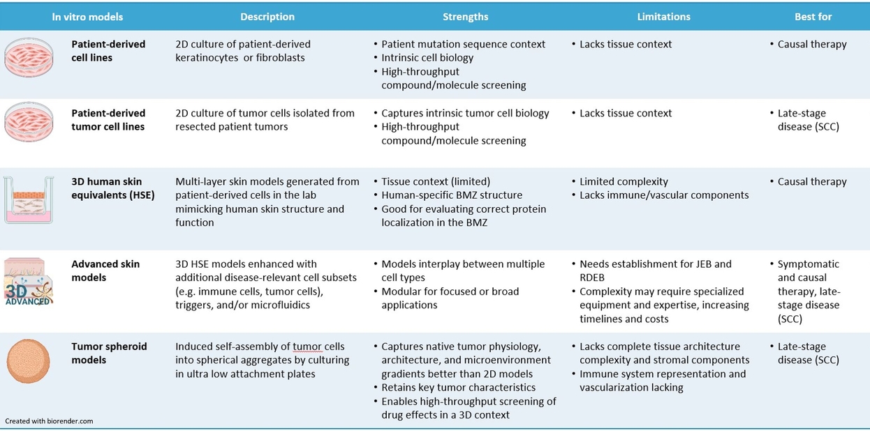
2D Cell Culture Models
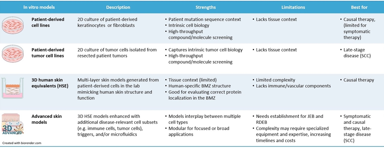
- Patient-derived cell lines (primary and immortalized):
Keratinocytes and fibroblasts isolated from patient skin biopsies that retain the causal genetic defect and loss of the affected protein. These models provide a reliable platform for mechanistic studies and preclinical evaluation. - Patient-derived tumor cell lines:
Tumor cells established from resected patient samples, enabling the study of tumor biology and the assessment of candidate therapies.
3D In Vitro Models
- Human Skin Equivalent (HSE) models:
Fully stratified human skin reconstructed in vitro from patient-derived cells. These models preserve the native mutation context and are well suited for causal therapeutic approaches, including gene editing, readthrough strategies, and antisense oligonucleotide-based therapies. Key readouts include protein restoration and resistance to blistering. - Advanced skin models:
Enhanced HSE systems incorporating disease-relevant cells, such as immune or tumor cells, and external triggers, such as Staphylococcus aureus infection. These models enable the analysis of inflammation, fibrosis, cellular activation, and early wound repair. - Tumor spheroid models:
Three-dimensional tumor aggregates generated by patient-derived cells that recapitulate tumor architecture and microenvironment. These systems provide a more physiologically relevant platform for preclinical therapy testing.
JEB in vitro models
JEB patient-derived keratinocyte and 3D HSE models accelerate gene therapy development by enabling evaluation of gene editing and gene replacement strategies, including CRISPR-based correction of COL17A1 and LAMB3, with functional readouts of protein restoration and blister resistance.
Advanced HSE models incorporating disease-relevant immune cells further capture disease-relevant responses, including inflammation, fibrosis, cell activation, and early wound repair.
RDEB in vitro models
RDEB models based on patient-derived keratinocytes and 3D organotypic skin equivalents support the development of gene therapies. They enable evaluation of gene editing, gene replacement, and readthrough approaches, with functional readouts including protein restoration and improved blister resistance.
Advanced 3D HSE models incorporating disease-relevant cell subsets, such as immune cells, further allow assessment of inflammatory and fibrotic marker expression, cell activation, and early wound repair.